Simple Model Recreates Hair Follicle Patterns in Two Mammal Species
A University of Geneva model shows hair follicle patterns can arise from cells following chemical signals. The PNAS paper covers mice and spiny mice.
A mathematical model in which skin cells follow a chemical signal reproduces the layout of hair follicles in two mammal species, researchers at the University of Geneva (UNIGE) report in the journal PNAS. The team says the pattern needs no detailed instructions for each follicle. Follicles appear on their own from local interactions between cells and chemicals.
The result challenges the explanation that has dominated for laboratory mice. The same mechanism also matched the unusually regular, strongly aligned follicle rows of the spiny mouse, which the older explanation cannot account for.
How hair follicles start
Long before birth, a mammal’s skin marks the spots where hair will grow. Hair, feathers and most scales begin as small structures called placodes in the embryo, and their arrangement differs from species to species. Scientists have spent decades asking what decides where each placode forms.
The older explanation for mouse hair
For laboratory mice (Mus musculus), the leading account is the expansion-induction model. A new placode releases a chemical that stops other placodes from forming too close to it. As the embryo’s skin grows and stretches, some areas end up far enough from existing placodes to escape that effect, and new placodes form there. The pattern fills in step by step.
The idea is widely accepted. According to UNIGE, though, scientists have not shown experimentally that it can explain the variety of follicle patterns across species.
Cells that move toward a chemical
Athanasia Tzika, a senior lecturer, and Michel Milinkovitch, a professor, both in the Department of Genetics and Evolution at UNIGE’s Faculty of Science, led a team that tested a different process called chemotaxis. In chemotaxis, cells move along differences in chemical concentration. White blood cells use it to travel toward areas of inflammation.
The researchers built a model of mobile cells in the dermis that respond to an attracting chemical released by the epidermis, the skin’s outer layer. They then simulated the growth of embryonic skin to see whether placodes would appear gradually. The simulation recreated the development seen in laboratory mice, including the same order of follicle formation that the expansion-induction theory had been used to explain.
“Placodes emerge spontaneously from local interactions between cells and chemical signals,” said researchers Muhamet Ibrahimi and Ebrahim Jahanbakhsh.
A second species with a different coat
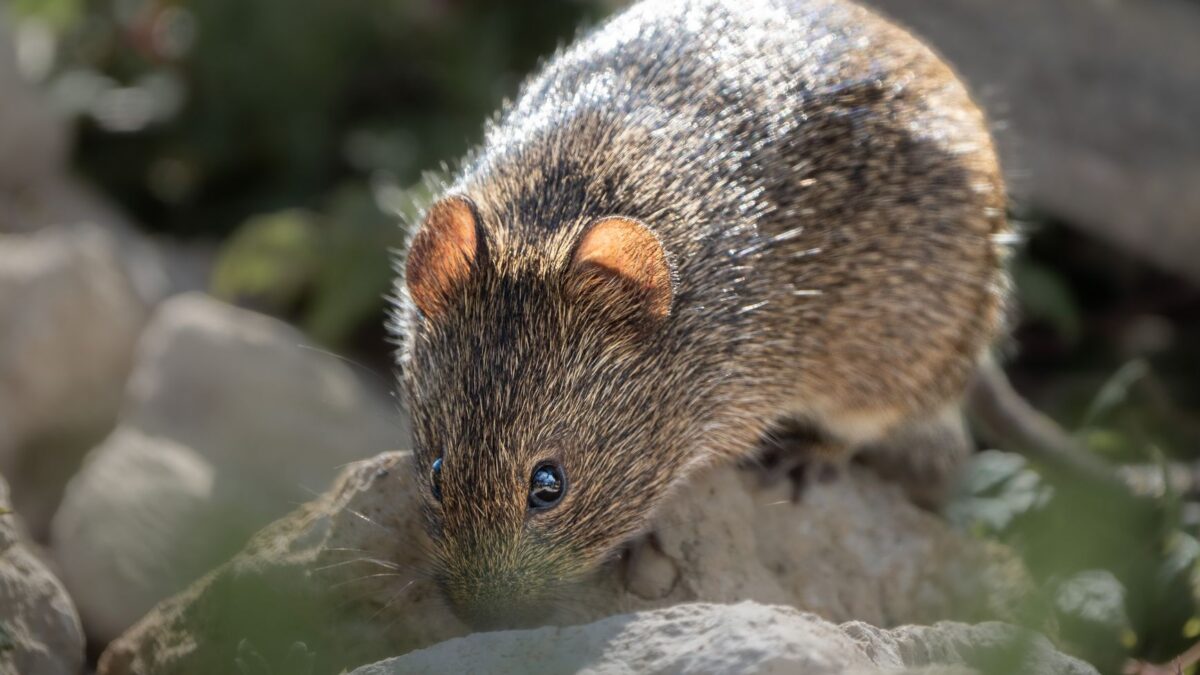
The team then tried the spiny mouse (Acomys dimidiatus). Its follicles form in an unusually regular pattern with strong directional alignment. The researchers adapted the simulation using information from three-dimensional images of developing embryonic skin, reflecting the species’ biochemistry and the way its skin grows.
The same process of cells responding to chemical signals reproduced the spiny mouse pattern. According to the team, the differences between the two species come from variations in cell interactions, chemical properties and skin growth, not from entirely different mechanisms.
What is still unclear
The study is a model built on experimental data and tested against two species. The authors word their conclusions as suggestions: that elaborate structures can emerge from basic interactions between neighboring cells, and that the same rules may apply more widely. Feathers and scales also begin as placodes, and the team says its finding could help explain that wider phenomenon, but the model was run only on mouse and spiny mouse skin.
Why it matters
If the mechanism holds up, one self-organizing process with adjustable settings could produce very different follicle layouts without a separate blueprint for each species. Tzika said the work suggests that simple cellular interactions can generate the diversity of tissue architectures seen across evolution, and that species differ in the interactions between cells and chemical signals rather than in the underlying process.
The paper is “Chemotactic self-organization captures the dynamics of mammalian hair follicle patterning” by Ibrahimi, Jahanbakhsh, Tzika and Milinkovitch, published in Proceedings of the National Academy of Sciences (DOI 10.1073/pnas.2530407123).
Source: ScienceDaily