Sleep Disorders Tied to a Smaller Thalamus in 57 Brain Imaging Studies
A review of 57 brain imaging studies links insomnia, sleep apnea and sleepwalking to changes in attention and decision areas. What the study can and cannot show.
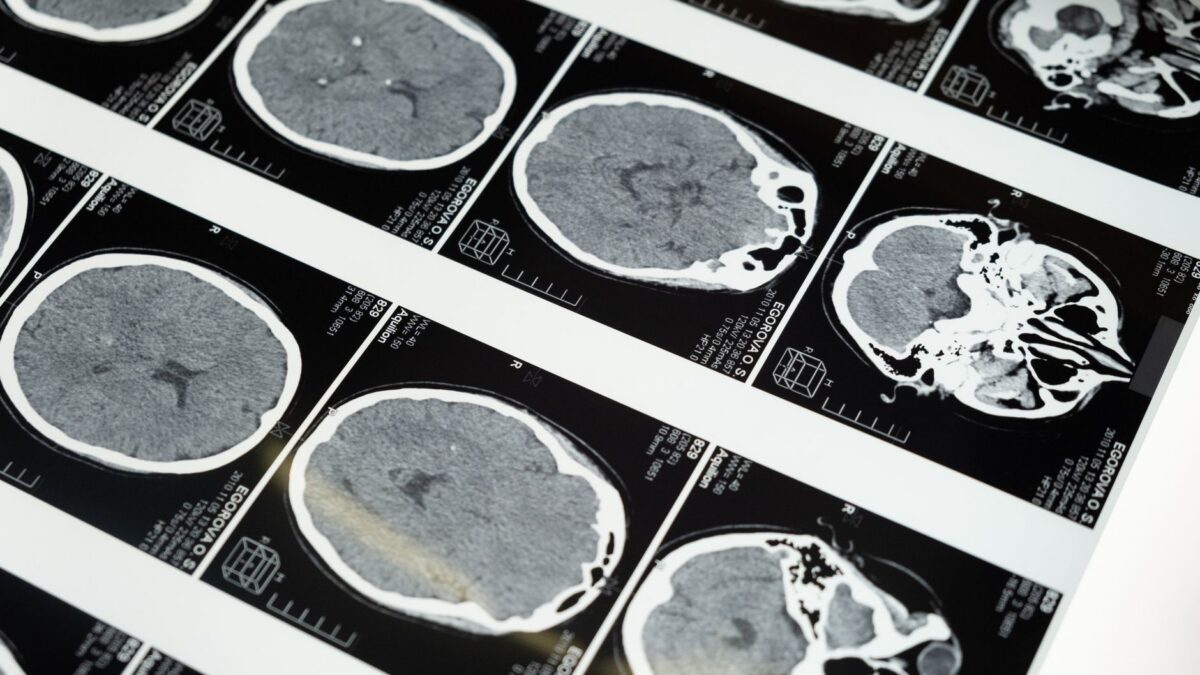
Adults with sleep disorders show structural differences in brain regions that handle attention, motivation and decision-making, according to a meta-analysis by researchers at Florida International University published in the journal Scientific Reports. The team pooled results from 57 brain imaging studies and found that both broad groups of sleep disorders were linked to reductions in the thalamus, a deep brain structure that helps process information and hold attention.
The study shows an association only. The authors say it cannot tell whether poor sleep changes the brain, whether existing brain differences lead to sleep problems, or whether both happen.
How the FIU team compared sleep disorders
Most research examines one sleep disorder at a time. The group at FIU’s Center for Children and Families instead combined imaging studies of adults and compared several disorders side by side, looking for changes they share and changes unique to one type.
They sorted the conditions into two groups. Dyssomnias, such as insomnia and sleep apnea, make it hard to fall asleep, stay asleep or get restful sleep. Parasomnias, including sleepwalking, nightmare disorder and sleep terrors, involve unusual events or behavior that break up the normal sleep cycle.
A shared change in the thalamus
Both groups showed reductions in the thalamus, and the researchers traced the change to a part of it called the pulvinar. The pulvinar helps steer attention toward what matters and supports cognitive control, the ability to manage thoughts and actions while doing a task.
These areas connect to larger networks used for concentrating, making decisions and reacting to surroundings. The authors suggest that differences there may help explain why people with sleep disorders often react more slowly, struggle with decisions and make more errors or have more accidents.
Lead author Katharine Crooks, who recently completed a doctorate in cognitive neuroscience at FIU, said most research looks at sleep disorders one at a time and that pooling studies revealed patterns they share.
Extra differences in sleepwalking and nightmares
People with parasomnias also showed changes in the posterior cingulate cortex, a region involved in motivation, decisions and emotional regulation. The researchers say this may help explain why sleepwalking, recurring nightmares and sleep terrors sometimes come with problems in mood, behavior or emotional control.
The analysis found no consistent structural change seen only in the dyssomnia group, which includes insomnia and sleep apnea.
Why it matters
An estimated 50 million to 70 million Americans have a sleep disorder, according to the FIU release. Knowing which brain regions are involved in which disorders could, the authors say, eventually help doctors spot sleep problems earlier and design treatments aimed at the specific brain systems affected.
Senior author Matthew Sutherland, a cognitive neuroscientist at FIU, said combining many studies gives “a clearer picture” of how sleep disorders relate to brain structure and function. The findings come from earlier imaging work rather than a new trial, and they describe group differences, not a test for any one person.
What comes next
The FIU team plans to study individual parasomnias in more detail, including sleepwalking and REM sleep behavior disorder, to work out how each one relates to changes in the brain.
Source: ScienceDaily