Alzheimer’s Shrinks Two Brain Regions Before the Hippocampus
Johns Hopkins scans show the entorhinal cortex and amygdala lose tissue early in Alzheimer's, maybe a decade before memory loss. Methods and limits inside.
Two small brain regions next to the hippocampus lose tissue earlier in Alzheimer’s disease than the hippocampus itself, and the change may show up as much as a decade before memory problems begin, according to researchers at Johns Hopkins University. The regions are the entorhinal cortex and the amygdala.
Doctors who look for Alzheimer’s damage on brain scans usually focus on the hippocampus. The new work suggests that measuring the two neighboring regions together could flag people at risk sooner. The study appeared in NeuroImage Reports (DOI 10.1016/j.ynirp.2026.100392) and is being presented at Neuroscience 2026, the annual meeting of the Society for Neuroscience, Johns Hopkins said in a release published Nov. 14.
How the team tracked early shrinkage
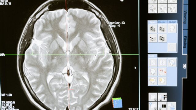
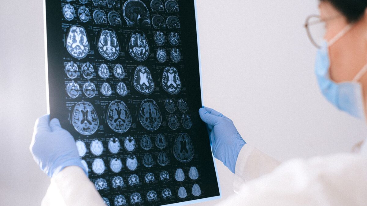
The scientists went through hundreds of MRI and PET scans. MRI shows the shape and size of brain structures, and PET can reveal certain proteins. The scans came from two separate research projects: the Alzheimer’s Disease Neuroimaging Initiative and the Biomarkers of Cognitive Decline Among Normal Individuals study.
Both projects followed people at different stages of the disease over time. Some had no symptoms, some had cognitive impairment, and some were patients who later died of Alzheimer’s. In both groups the same pattern turned up. In the early stages, the entorhinal cortex and amygdala showed greater relative tissue loss than the hippocampus.
Each region has its own job. The entorhinal cortex helps the brain pull up memories that were already stored. The amygdala takes part in emotional memory, shaping how emotional experiences are processed and remembered.
Shrinkage overlaps with tau buildup
The researchers also compared the tissue loss with tau, a protein closely tied to Alzheimer’s. In a healthy brain, tau helps hold the inner structure of nerve cells in place. In Alzheimer’s it can pile up abnormally and twist into tangles that disrupt normal brain function.
Using PET imaging and digital pathology, which analyzes tissue samples with computer tools, the team found that areas where gray matter shrank matched areas with tau-related changes. Gray matter holds large numbers of nerve cell bodies and does much of the brain’s information processing. That overlap makes the two regions stronger candidates as early markers of the disease.
Senior author Michael Miller, a biomedical engineer, said the brain changes over “at least a 5-to-10-year” stretch before symptoms. He said treating people at the 2, 3 or 4-year point would be far better than waiting 15 years until memory symptoms show.
What it means for spotting Alzheimer’s earlier
Alzheimer’s is the most common cause of dementia worldwide, and its changes build up in the brain for years before people notice forgetfulness or confusion. Researchers see that quiet period as a window in which treatment might protect more of the brain, but using it requires biomarkers: measurable biological changes that show a disease is present or getting worse.
Co-author Marilyn Albert, director of the Johns Hopkins Alzheimer’s Disease Research Center, said the aim is to treat people before symptoms develop. Co-author Meaghan Morris, a neuropathologist, said the approach could help sort people by risk and identify who is likely to have cognitive decline soon. As brain imaging gets cheaper and easier to reach, the researchers picture doctors checking these regions in people with a family history or other risk factors as early as their 30s or 40s.
What the study does not yet show
The findings point to possible early biomarkers. They do not amount to a clinical test that can predict Alzheimer’s years in advance, and more research is needed before doctors could rely on these changes for diagnosis. The work is based on scans and tissue analysis; no treatment was tested.
Next, the team plans to study the two regions at a much smaller scale, looking at the molecular changes around abnormal tau and how they drive tissue loss. Miller said that once researchers understand these “molecular neighborhoods,” they can work on therapies.
Source: ScienceDaily